News Story
New system could improve thermal devices used to check our temperatures and respiration
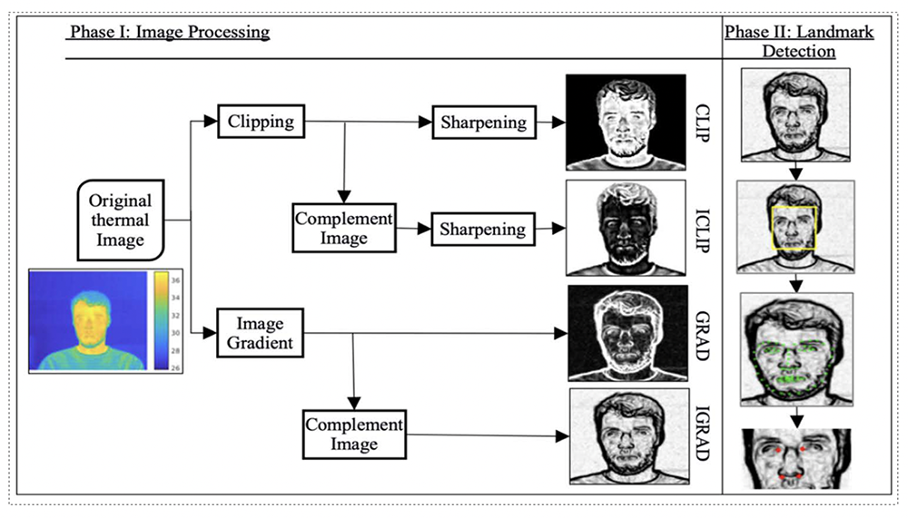
CLICK FOR LARGER IMAGE. System framework for two-phase inner canthus and outer-nostril edge detection. (Fig. 1 from the paper)
During the COVID-19 pandemic, we all got used to being remotely scanned by thermal camera devices that remotely checked our temperature and respiratory rate. These infrared thermographs (IRTs) were widely installed in medical facilities, airports and offices, and are likely here to stay because the vital signs they measure are useful infectious disease biomarkers.
To accurately estimate body temperature and respiration rate based on thermal images, it is essential to ascertain the optimal locations in the images for signal analysis. Sometimes it takes some time as we move our heads around to help the IRT actually find what it is looking for—an area around the eyes called "inner canthi" for body temperature and a different area around the nostrils for respiratory rate.
Hoping to make the process faster and more efficient, researchers are improving the software algorithms that automatically identify the best facial locations to extract these vital signs. It’s actually pretty difficult to automatically identify these regions in thermal images—harder than it is, for example, in regular photos, which are rich with exploitable features.
Now ISR-affiliated Professor Min Wu (ECE/UMIACS) and her colleagues have developed a unique system that can detect inner canthi and outer nostril edges directly in thermal images in two phases.
Detecting Essential Landmarks Directly in Thermal Images for Remote Body Temperature and Respiratory Rate Measurement With a Two-Phase System has been published in IEEE Access.
The authors are Wu; her Ph.D. student Zachary McBride Lazri; two of her alums—Qiang Zhu (ECE Ph.D. 2020) and Mingliang Chen (ECE Ph.D. 2021), both now research scientists at Meta; and Quanzeng Wang of the Center for Devices and Radiological Health at the U.S. Food and Drug Administration.
In Phase I of the system, original thermal images are processed in four different ways to enhance facial features and facilitate inner canthus and nostril detection. In Phase II, landmarks of the inner canthi and outer nostril edges are detected in two steps: (1) face detection using Single Shot Multibox Detector (SSD) and (2) facial landmark detection to locate the inner canthi and outer nostril edges.
The system automatically and accurately estimates the inner canthus and nostril locations in thermal images. It can be applied in IRT algorithms to provide reliable temperature respiratory rate estimates during infectious disease outbreaks.
Published August 26, 2022
Related Stories
Stories / January 13, 2022
Connected autonomous vehicles hold promise for alleviating...
Stories / July 30, 2021
Derek Paley's e-scooter work featured in Washington Post

Stories / December 8, 2020
‘Smellicopter’ drone uses live moth antenna to seek smells,...
Stories / August 24, 2020
Wu, Milton receive NSF funding to improve telemedicine

Stories / November 5, 2019
ISR, ECE, CS, UMIACS faculty present 12 talks at Northrop...

Stories / August 27, 2019
ISR remembers Carlos Berenstein

Stories / July 19, 2019
Narayan is PI for NSF information-theoretic signal processing...

Stories / September 3, 2016
Alumna Jing Yang begins tenure-track position at Penn State

Stories / December 7, 2023
‘Priming’ helps the brain understand language even with...
Stories / November 29, 2023
Alum Fumin Zhang elected to IEEE Fellow